
Cell Quality Control (CQC)
The identification and counting of cells, including discrimination between living and dead cells, is an important component of any Quality Control procedure for cell-based manufacturing. Traditional methods for cell viability assessment use fluorescent dyes or labels to differentiate live cells from dead, but are destructive to cell samples while also adding complexity, time, and risks to the process.
Ovizio’s patented double differential digital holographic microscopy (D3HM) circumvents the issues surrounding traditional methods by capturing images of cells without the need for dyes or labels. D3HM also provides additional cell parameter data that cannot be obtained with other cell counting technologies, thus generating a unique holographic fingerprint for every cell imaged.
Individual cell fingerprints are fed into our proprietary OsOne machine learning software, which automatically discriminates between living and dead cells, counts them, and gives access to in-depth quality attributes and dynamic properties of your samples.
Suspension cells
You will be informed about your cells’ Critical Quality Attributes (CQAs) such as viability, total cell density and viable cell density, aggregate rate for cells in a range from 4 μm to 150 μm (depends on magnification, cell line); without any dye or labels. Living and dead cells have significant holographic patterns that can be distinguished and accurately counted.
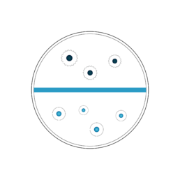
Images of CHO (Chinese hamster ovary) cells with cell detection and viability assessment mask overlay
Key: green dots – viable cells; red dots – dead cells; yellow dots – aggregates.
The Ovizio cell counts and viability measures are often compared with measures from a reference method (offline cell counter such as ViCell, Nucleocounter, etc. using dyes or labels; or a flow cytometer).
Case studies
For more comparison studies, download our posters:
Adherent cells
Ovizio’s qMod quantitative camera is a phase contrast camera that can convert a regular brightfield microscope into a versatile 3D quantitative imaging platform for biological applications. It can be attached onto the C-mount where a camera is usually attached to the microscope. The qMod is a convenient and fast solution for direct cell counting in transparent supports, from petri dishes, flasks and multi-well plates to multi-layer vessels.
This provides users with access to the D3HM technology, allowing them to take advantage of the dye-free setup to monitor cell viability, cell confluency, cell morphology. The throughput can be increased by connecting the qMod camera to a microscope with a motorized stage. The image acquisition & computation is done in less than 3 seconds and, thanks to our technology, a post-acquisition refocusing can be done.
In the pictures below, MRC5 & VERO cell lines are cultivated in T75 flasks. Image acquisition was performed with the qMod coupled to Olympus or Leica microscopes.
As described in this picture, cell confluence can be observed and accurately measured in a dye-free setup.
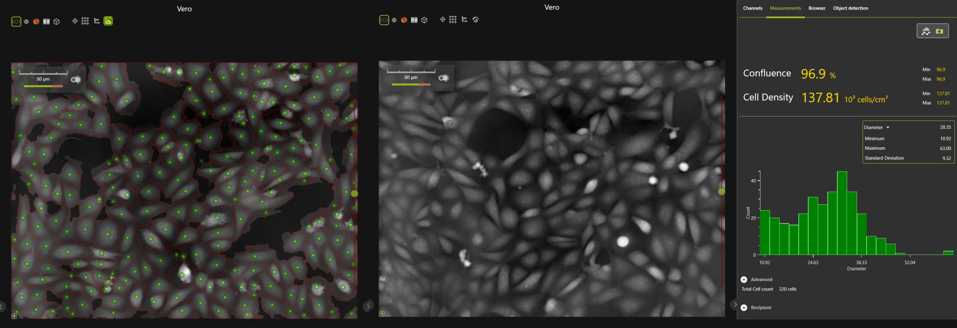
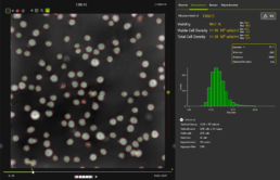
VERO cell line – confluence measurement
Phase images of VERO cells in a T75 flask, without (left) and with cell segmentation (right). Each cell is outlined with a red line and the green spots highlight the viable cells. The cell confluence is measured at 83.7% with a cell density of 102.49×10³ cells/mm2. Both pictures are screen shots from the OsOne software.

MRC5 cell line – confluence measurement
Phase images of MRC5 cells in a T75 flask, with (left) and without (right) cell segmentation. Each cell is outlined with a grey area. The cell confluence is measured at 47.6%.
Both pictures are screenshots from the OsOne software.
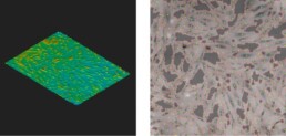
Human Meschenchymal Stem Cells (Hu MSCs)
in Xpansion® bioreactor (Pall Lifesciences)
3D histogram imaging (left) and phase image (right) of MSC cells and automated cell detection. Each cell is outlined with a red line and the green spots highlight the viable cell. Dark grey zones indicate non-colonized surface. Here, the confluence is measured at 70%.
Need any technical help?
Please email us at support@ovizio.com
T: +32 2 600 50 90
E: info@ovizio.com
Ovizio Imaging Systems NV/SA
Av. Kersbeek 306
1180 Uccle / Belgium
Website by Monkeys not Donkeys