
T-cell activation

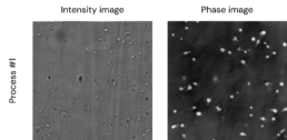
Our iLine F automated microscope provides real-time tracking, in the bioreactor, of the morphological changes associated with T-cell activation, without interrupting the expansion process. Our double differential digital holography technology does not require dye or labeling, allowing the cells to continue their growth process unhindered.
T-cell receptor engagement, with accompanying costimulatory signals, control the level of activation and functional potential of individual T cells. This can be further manipulated, ex vivo, via antigen-presenting cells (APCs) loaded with the desired antigen, dendritic cells (DCs) or anti-CD3/anti-CD28 beads, triggering a physiological activation and expansion of human T cells for potential clinical applications.
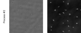
The changes in T-cell morphology that occur during immunological synapse formation are essential for T-cell activation. T cells have been shown to change from a spherical to elongated morphology, which is believed to be characteristic of mobile T cells scanning or spreading towards APCs, DCs or reacting to any compound that will target the T cell receptor and its costimulatory molecule, like anti-CD3/anti-CD28 beads.
This new elongated and enlarged T-cell morphology can be tracked in real time in a bioreactor without disrupting the expansion process, using our iLine F automated microscope. Thanks to our double differential digital holography technology, no dye or labeling is required, allowing the cells to continue their growth process without any detrimental consequences for the culture.
The OsOne software and its associated machine learning platform provide a quantitative measurement of these activated T cells: both a total count and percentage of activated T cells in the culture. Therefore, we offer a continuous quality control of the culture with no risk of contamination.
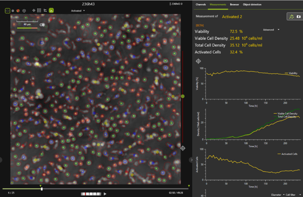
In this phase image, analyzed and segmented by the OsOne software, several populations have been tagged within a single sample:
Key: green dots – viable cells; red dots – dead cells; Blue dots: activated cells; Yellow dots: cell clusters.
Case studies
Need any technical help?
Please email us at support@ovizio.com
T: +32 2 600 50 90
E: info@ovizio.com
Ovizio Imaging Systems NV/SA
Av. Kersbeek 306
1180 Uccle / Belgium
Website by Monkeys not Donkeys